


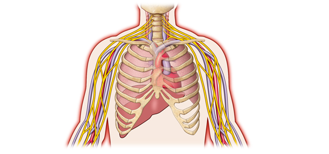
Thorax: Thoracic Outlet Syndrome (TOS)
MRI, MRA, and MRV. A two- and three-dimensional image reconstruction, with applications including evaluation and diagnosis of thoracic outlet syndrome (TOS).
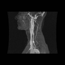

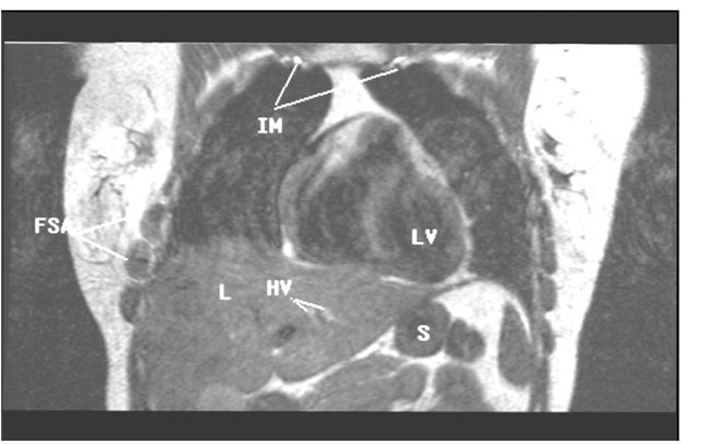
Figure 8: This is an anterior image of the coronal abduction external rotation of the upper extremities sequence in Figure 7. This image displays high signal intensity (white) internal mammary (IM) and hepatic veins (H) reflecting increased thoracic and abdominal pressure secondary to decreased venous return on a T-1 sequence. First fascicle of the serratus anterior muscle (FSA), liver (L), left ventricle (LV), stomach (S). With permission from James D. Collins, MD. Read complete article here.
More...
brachial_plexus_compromising.pdf
tosinfo.com
All rights reserved.